
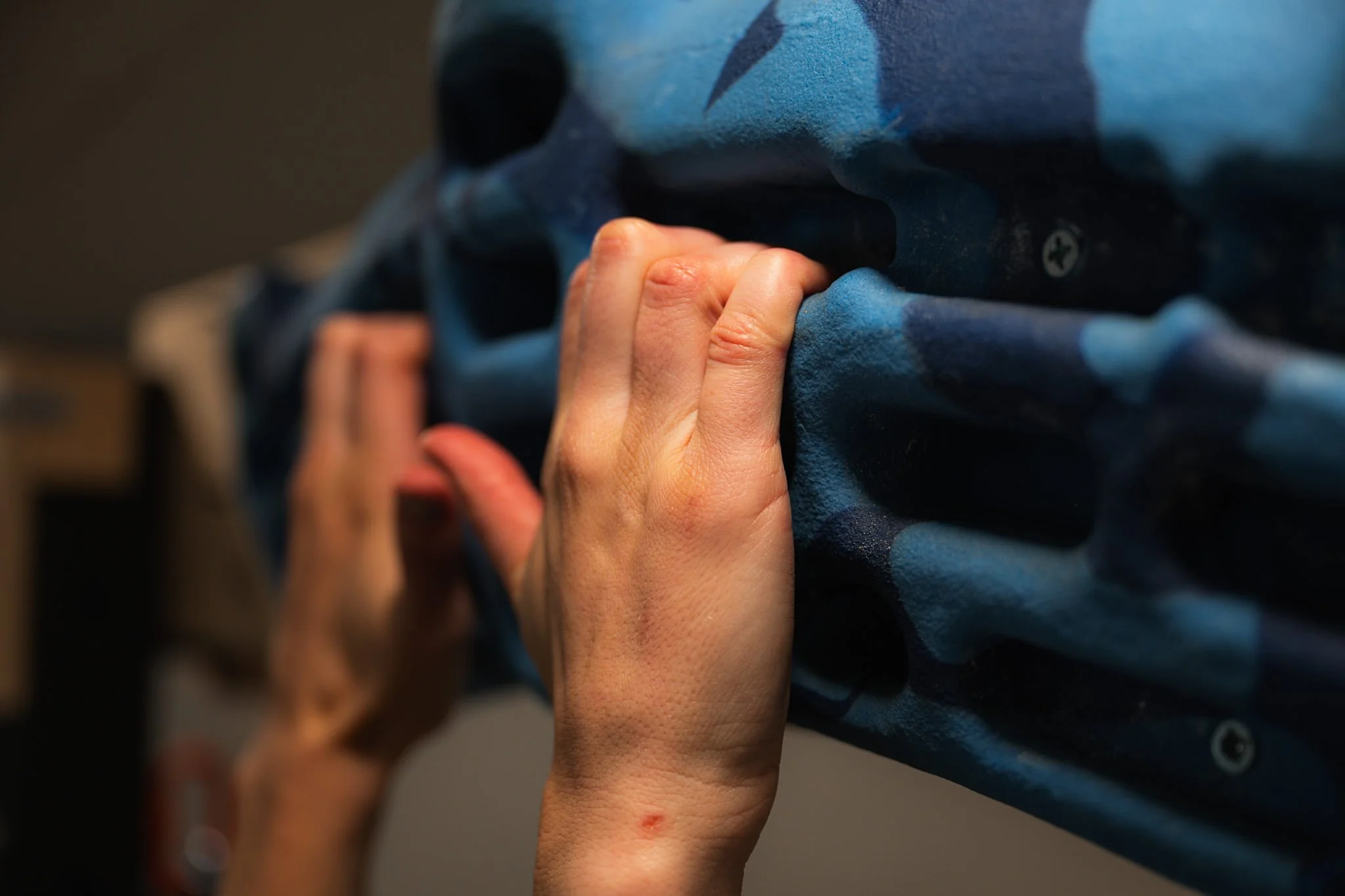
Diagnostic Ultrasound for Finger Injuries
Finger injuries make up more than 50% of rock climbing injuries¹, and ultrasound imaging is a valuable tool when it comes to aiding in the diagnosis of those injuries.
Though a key portion of a physical therapy exam is always based around clinical tests and measurements, ultrasound imaging is a safe, inexpensive way to shed additional light on the severity of an injury and specific tissues involved.
Point-of-care ultrasound can be done in real-time, in the physical therapy clinic, at no additional cost.
-
Diagnostic ultrasound uses sound waves to create an image of tissues. It's most familiar to many folks for its use during pregnancy, but it can also be used to visualize other tissues, including muscles, tendons, and ligaments.
-
Yes! Ultrasound has no ionizing radiation (unlike x-rays). And compared to MRI, it is much less expensive, is quicker, can be done right in the clinic (no referral), and can be used for side-to-side comparisons and to look at real-time movement.
-
Diagnostic ultrasound is highly reliable for small, superficial structures (like those in the fingers), and is considered the first-choice imaging option for finger injuries like pulley tears/sprains, tenosynovitis, and others.² ³
Diagnostic Ultrasound FAQs
Diagnostic ultrasound is offered as part of in-person physical therapy sessions for finger injuries at the clinic in North Laurel, MD.
Cole K, Uhl R, Rosenbaum A. Comprehensive Review of Rock Climbing Injuries. J Am Aced Orthop Surg 2020;00:1-9.
Rossi F, Romano N, Martinoli C, et al. Wrist and Hand Ultrasound: Reliability of Side-to-Side Comparisons of Very Small (<2-mm) Clinically Relevant Anatomic Structures. J Ultrasound Med. 2018;37(12):2785-2795.
Schoffl V, Schoffl I, Frank L, et al. Tendon Injuries in the Hands in Rock Climbers:Epidemiology, Anatomy, Biomechanics and Treatment -An Update. Muscle, Ligaments andTendons Journal2020;10 (2):233-243.